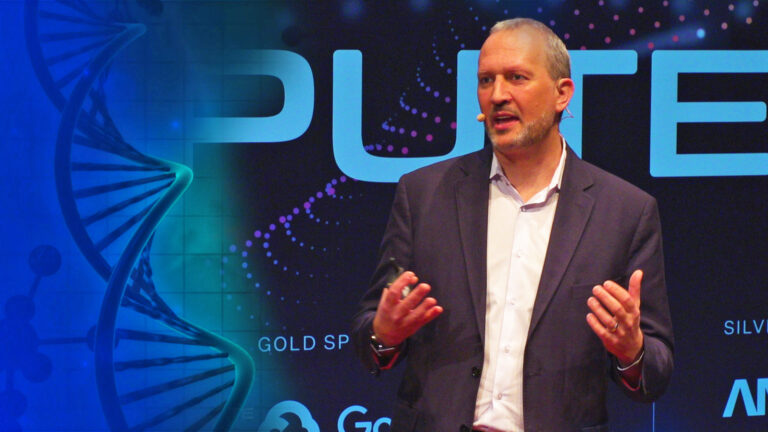
Accelerating Medicine with Modern Genomics
Speaker:
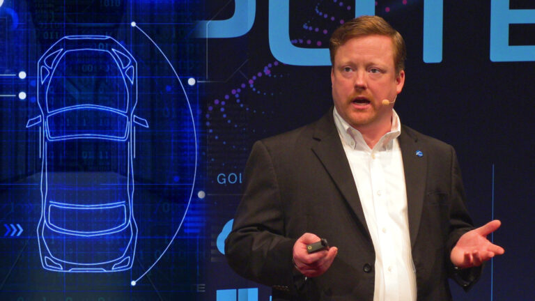
Please welcome to the stage, the COO of Bionano Genomics, Mark Oldakowski.
Mark:
Thank you for all of you being here. Thank you to Rescale for getting us together to talk about big compute and HPC. I’m going to start off this morning with a little bit of audience participation. It’s a little bit bright for me here, so no hands with applause. I want to know who here has heard of the terms precision medicine or personalized medicine? All right. Good. You were paying attention to Joris talk because he mentioned one of those terms. Now, we use those terms in life science to explain a vision that we have.
Mark:
That vision is to find the unique signature of the disease that’s ailing you, the unique genomic signature and to create a therapy that addresses that signature very precisely but also takes into account your biological uniqueness. We want to do that to maximize efficacy of that therapy and to reduce the side effects, laudable goals.
Mark:
Over the next 20 minutes, I’ll share with you that that is an aspirational goal, that we’re not there yet. We have some hints of it and I’ll share with you some of those, but we are not there yet. One of the critical tools that’s required for us to get there, to realize that vision is big compute, high performance computing. I’m sure many of you have heard a lot of good news about DNA sequencing being applied to medicine. There’s been a lot of buzz around that in media, rightfully so, but would you believe that the most prevalent clinical testing that’s occurring for complex diseases worldwide is actually test that was brought to the clinic over 60 years ago.
Mark:
That requires a little bit of innovation to get past that. I’ll tell you why. One of these tests is called karyotyping and with karyotyping, one gathers all of the chromosomes from a cell and stains those in some unique patterns and then looks at those chromosomes with a microscope. Different kinds of microscopes. Here’s one, but what you end up getting is these 46 chromosomes that have the stain pattern. There’s software that helps you orient those, so that they all are presented for your viewing pleasure in the same way, but then a very trained side of geneticist looks at those chromosomes, looks at those patterns and tries to determine whether they’re normal or not normal. Now, the resolution at which they are looking at is 10 million base pair. Base pair is a unit of measure of DNA. Their resolution is 10 million base pairs, right?
Mark:
If an event, a rearrangement occurs at a lower level than 10 million, they will not see it. If it’s a complex set of rearrangements, which I’ll show you in a few minutes, they are likely to miss it. Those are important considerations because these are the folks that are telling the doctor what specific disease you may have, and these are the folks that help to provide data to pharmaceutical companies to find new drug targets.
Mark:
All right. I’ll show you a few karyotypes. We’ll step through a few of them. Before we do, let’s take a look at what a normal karyotype looks like. All right. You see 23 pairs of chromosomes with the last two being your sex chromosomes. Two X’s determine that you’re a female. XY, you’re biological male. All the other chromosomes are in pairs and you get one copy from mom, one copy from dad. All of this is contained in the single cell on each chromosome. The chromosome is made up of DNA. Your entire genome is represented on these 46 chromosomes.
Mark:
They’re laid out in the way where the larger chromosomes, the ones that contain more DNA are first and the ones that have less are last. In this representation, chromosomes one have far more DNA and chromosome 22. Within that DNA, there are regions that encode for genes and genes drive action in the body, drive proteins. All right. The other thing to note is that the pairs are about the same length, right? Chromosome one, those two chromosomes should be about the same length. I’m using very precise terms here because that’s how precise this technique is. All right. Let’s look at our first abnormal chromosome. This is a disease chromosome. Can everybody spot the difference? Right?
Mark:
Chromosome 21, there’s three chromosomes in that grouping. This is called trisomy 21, also referred to as down syndrome. It is a disease that’s often tested for or regularly tested for in the prenatal clinic. All right. Great. That precision, you guys are all budding cited geneticist. What else do we see here? Do we see any other anomalies? Chromosome one, both of those chromosomes look about the same. Everything else looks about the same. It’s an XY male, right? Okay. Things look okay, so I’m happy with my diagnosis. I write it in the standard notation that’s been around for decades. I write 47. I see 47 chromosomes, XY, XY male, plus 21. There’s an extra chromosome, 21 and I hand that to the doctor and done. Great. All right.
Mark:
How about this one? A lot of weirdness going on. If you’re trying to look for any short chromosomes, meaning short meaning DNA is missing. Now, you look at chromosome four, chromosome six is that just… Are those pairs shorter or is it just an optical illusion? I don’t know. I’m not a cited geneticist, but one thing that is clear is that we’ve got this O. We call that a ring chromosome. Somehow, the two ends of that chromosome got fused together during the DNA replication process and created this O chromosome, which is pretty detrimental to the patient that introduces microcephaly. Small brain, developmental delay, short stature among other things. Pretty bad to have a ring chromosome on chromosome 15, but fairly easy to pick up, right? We picked it up.
Mark:
I think you all would say, “Boy, what’s wrong with karyotyping? This is easy. We could do that. We can write software to pick these things up.” Right? All right. Well, let’s look at something more complex and this is where things start falling apart for this method. What’s going on here? We’ve got a lot of errors pointing to a lot of things. Way too many events to mention, but we obviously see missing chromosomes, right? Chromosome 12 and 16 and 20 don’t have a mate. We see trisomy 22. We see chromosome five has got a shorter chromosome. A lot of genomic information is missing there. Chromosome 19 as well. Chromosome seven, I’m not sure what that’s pointing to, or maybe it’s some extra information on one of those chromosomes.
Mark:
Hey, it’s a female. At least we got that right, right? Two X’s. We also have these other things at the end, MR and AR. What the heck is that? Those aren’t typical chromosomes or they’re not chromosomes at all. They’re actually DNA that was leftover during the building process of chromosomes for the cells where the machinery gotten mixed up and didn’t know where to put it. They put it together and it created these new chromosomes. Boy, pretty complicated. Now, this is an actual signature for a myelodysplastic syndrome, which is a pretty serious blood disorder by itself, causes a lot of issues, but it also leads very often into a very aggressive form of leukemia, a blood cancer called acute myeloid leukemia. This is a signature of this MDS syndrome.
Mark:
I say a signature because there are different kinds of signatures for MDS, and they affect people in different way. Boy, as an engineer, when I look at this, a few things come to mind. One is, boy, it’s easy to miss something, right? There’s so many events going on here. It’s easy to miss something. Also, if I’m looking at a resolution of 10 megabase pairs, and I see these many events, how many events am I not seeing that are below that are hidden under the surface? Right? Are those also important to drive therapy and drive drug discovery?
Mark:
Boy, is this scalable? Right? Imagine the train side of geneticist that you would need to do this at full scale for millions of patients and do it not only once, but do it every week to follow patients during treatment. This is not scalable. You can’t make that work. Talk about therapies, now most genes are a hundred times smaller than the resolution that I can see here. If I see a rearrangement, disruption, missing information, to me, it looks like it’s impacting many, many genes, but which genes are actually impacted? Which ones, as a pharmaceutical company, do I go after and target, right? Which ones are the ones that are causing that disease? I don’t know. I don’t have that precision. That’s a problem. However, despite all of those issues, karyotyping is used all around the world as a primary way to diagnose complex disease.
Mark:
It’s used in all of the top… All of the cancer centers in the US because there is nothing better, 60-year-old technology. All right? Now, let’s imagine that in the modern genomics world, that this needs to look different, that we can take these chromosomes from cells. We can unwind the DNA. We can stretch it out predictably. We can mark it with mile markers, we know precisely where they are and then image it in high resolution and then have software do all of the identification of all the aberrations and provide those to the clinician.
Mark:
All right. This is what that looks like. What you’re seeing here is a high magnification image of actual DNA. Each of those strands is an actual DNA that’s been extracted from a cell and that DNA is confined in a nano fabricated device going through nanochannels that are 30 nanometers in width and those nanochannels allow a very fine confinement of that DNA where we know the dimensional properties and we can take measurements of very precise measurements between these various mile markers.
Mark:
From left to right, what you see in this video is that the DNA comes in, in its natural state. You see on the left, the DNA, it’s in a Gaussian coil and it’s unwound and stretched out in multiple steps along the way. Then, we take these images and these images look like the upper right hand where you have strings with beads on them. Right? Those beads are those markers that we put in, in very precise locations. We know where they are and we also know the distance between those markers because of our confinement of those DNA molecules with high precision. Then, software brings those molecules together, recreates the chromosomes, all 46 of them or more, if there are more.
Mark:
Then, the interesting things happen. The software is then able to look for rearrangements, aberrations, abnormalities in that DNA to gene level, sub gene level. All right. What do we do with this in practice? All right. One last karyotype. All right. This is a classic example of precision medicine. It’s arguably the first case of precision medicine that we’ve ever had. It involves the leukemia, chronic myeloid leukemia. It’s a leukemia that comes later in life in adulthood. The prevalence of that leukemia is increasing as our populations age.
Mark:
In 1959, two scientists were looking through hundreds of karyotypes. Poor souls, right? They were looking under a microscope, hundreds of these karyotypes. What they noticed is that for CML patients, that there was this particular signature that chromosome 22, one of the chromosomes was always shorter and chromosome nine was… One of those was a bit longer. They saw this in the majority of CML patients.
Mark:
Until 1973, all that was known as that boy, if you see this in the karyotype, then your patient most likely has CML. Okay. All right. Well, that’s better than nothing. Right? In 1973, there was another scientist that figured out the mechanism, right? She said, “It’s not just that nine has extra stuff from somewhere, and 22 has less DNA material. It’s actually an exchange of information.” Doing a DNA replication process and cells divide and grow, that chromosome nine and 22 exchanged information. Now, it wasn’t a fair trade. Nine got more DNA from 22, but 22 got something. What’s really key is that what happened on 22 was really revolutionary. Her discovery was that, “Boy, the material that 22 got included a gene that we now call ABL.
Mark:
It normally resides in chromosome nine. That gene got put on top of another gene on chromosome 22 called BCR. They got fused together, right? That fusion caused a new gene, right? That’s able to create a new protein that the body has never seen before. Now, that gene was always on, and that protein that had generated, its sole function was to keep itself alive on all costs, right? Cells are supposed to die. These cells were not dying, cancer. This was phenomenal. Armed with that knowledge, pharmaceutical company, Novartis developed the drug that they marketed in 2001 called Gleevec, and Gleevec’s sole purpose was to disrupt that protein, right? Target that protein disrupted, cell now dies its normal death. Patient cured. Phenomenal. Precision medicine at its best, right? It only took 42 years, but we got it.
Mark:
If I were to zoom in into both of those chromosomes because again, this is high resolution. We can zoom in really to a gene level. We can actually see that the precise location of the exchange of information created this BCR-ABL fusion gene. Okay? We can get this information from patient’s sample, from their bone marrow, to answer in the clinician’s hand in three days today and it’s getting faster.
Mark:
Now, what else do we see here? Boy, there’s a whole lot of dots and blue lines going on here. Additional exchange information. You see some sharing with chromosome 14. A lot of that is natural variation between individuals. We can filter that out by having and creating control databases. These control databases are databases that are based on ethnicities and so we can filter out many of those, but some of the things that would not get filtered out is these additional translocation events, chromosome 14. If one were to zoom in, where that exchange of information from chromosome 14 happens on 22 and nine, you’d see that in the same location as the BCR-ABL fusion. What that means is that this is a very rare three-way translocation, which is a novel signature to CML than the standard 922 signature. The reason that’s important is a novel signature can often mean different progression of that cancer, different resistance to the therapies. It’s another target for therapy.
Mark:
Is this better? It’s precise. It’s fast, and it gives insights to new drug targets that I can develop therapies for. Phenomenal. Phenomenal progress, but it’s not all done. Let me show you a more complex case. This is a variation plot, the circus plot of late stage ovarian cancer. You see a massive amount of rearrangement here. This is incredible. You see exchange of information from almost every chromosome to other chromosomes. You see things like in chromosome eight, which we call a chromothripsis event, which is through mechanisms that are not completely understood yet. Chromosome eight got shattered and then, the rebuild machinery didn’t know how to put it back together. It did the best it could, and you see a massive amounts of rearrangements within chromosome eight.
Mark:
This is ovarian cancer where we don’t have too many known drug targets here. We don’t really know which of these mutations we should be targeting. That’s the other problem. We have an effective and efficient method to discover these variations, but now we need to apply it to mass populations to help derive new targets, to help find new drug targets. We need to do this at scale.
Mark:
How do we intend to accelerate that and why do we need to accelerate it? Because we must. According to the National Cancer Institute, there are over 430 new cancers every year per 100,000 individuals. New ones every year, which are 160 individuals die of cancer every year per 100,000. This is a big scale problem. We need to provide more genomics data more quickly. The technologies like what Bionano has, we’re able to provide 10 times more data today than we were able to just 12 months ago. Ten times more data, which means that traditional computing just cannot scale with the amount of progress that we’re making and that rate of data generation is only increasing. It’s only getting faster.
Mark:
There’s more data, and the data is richer. We need HPC to be able to find these molecular signatures to figure out what to target with a therapy. We can’t do that with an on-prem HPC solution because you just can’t keep up. You just can’t scale fast enough. Solutions like what Rescale provides, allow us to really look at this in mass scale to really provide valuable intelligence to clinicians and therapy companies and hopefully bring precision medicine into the now as soon as we can. Thank you.